End of this page section.

Begin of page section: Contents:

Sample Preparation

The living sample
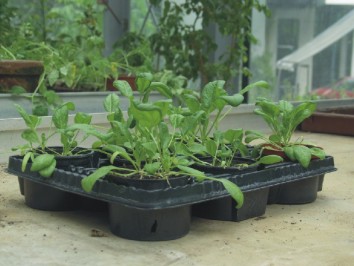
The living sample gets harvested and small pieces are fixed (chemical or cryo) and embedded in plastic.

Light microscopy
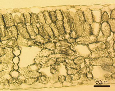
After the embedding semithin sections (1-5 µm) are getting observed for interesting features in the light microscope.
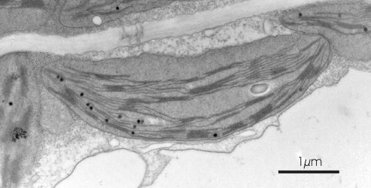
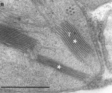
Electron microscopy

Ultrathin sections (50-90 nm) are made to investigate interesting features in the transmission electron microscope.

TEM-micrograph: thylakoid system in a chloroplast.

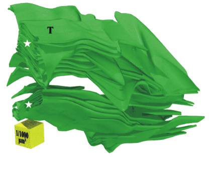
3D reconstruction

Series of ultrathin sections are reconstructed to a 3D image by computer programs.

More Infos

End of this page section.

Begin of page section: Additional information:

End of this page section.